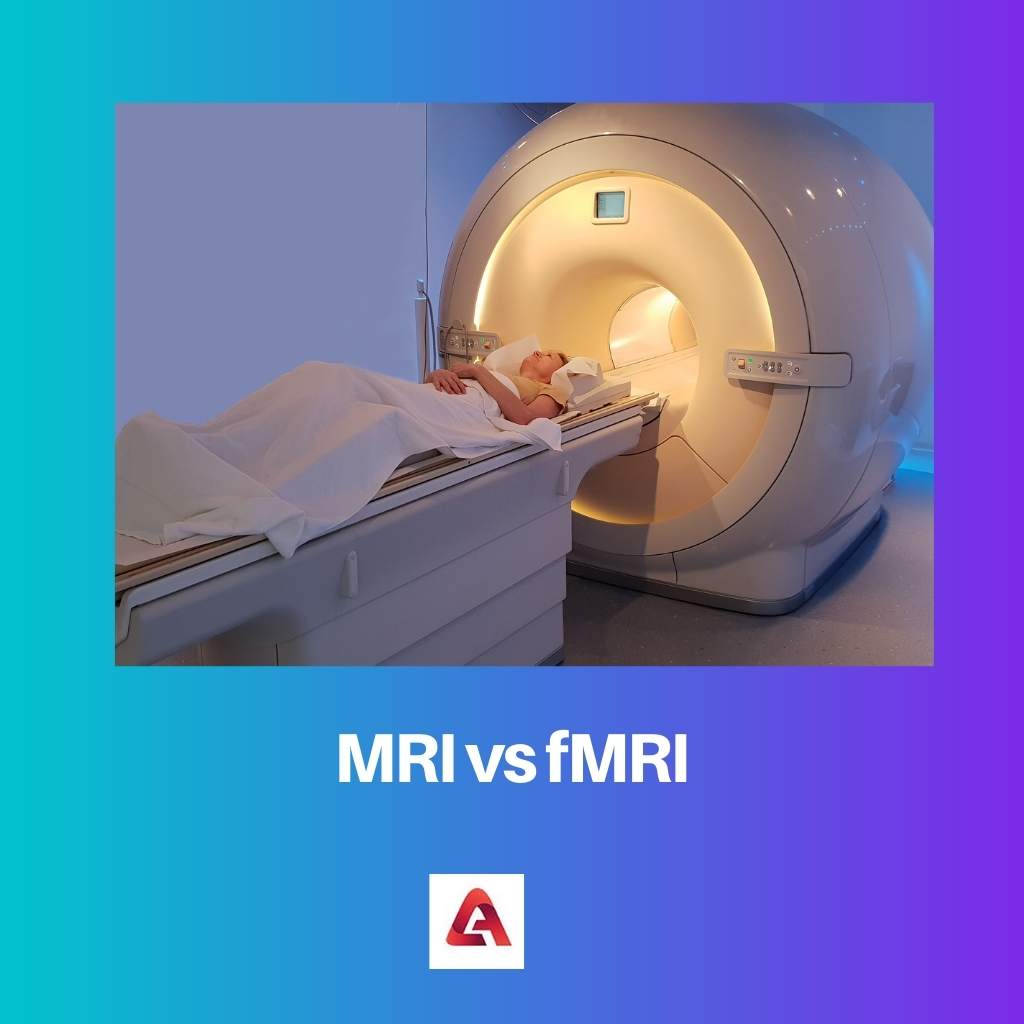
Eeg Vs. Mri Vs. Fmri , Electroencephalography functional magnetic resonance imaging
Di: Jacob
Concurrent TMS-EEG-fMRI opens an exciting noninvasive avenue of subject-tailored network research into dynamic cognitive circuits and their dysfunction. In MEG/EEG, you can follow brain activation with millisecond precision.Combining EEG and fMRI allows for integration of fine spatial and accurate temporal resolution yet presents numerous challenges, noticeably if performed in real .Geschätzte Lesezeit: 7 min
Electroencephalography functional magnetic resonance imaging
Designing studies: MEG vs fMRI. La risonanza magnetica viene utilizzata per diagnosticare condizioni .The combination of electroencephalography (EEG) with functional magnetic resonance imaging (fMRI) forms a powerful tool for the investigation of brain function, but concurrent .Functional MRI (fMRI) is widely applied in clinical and preclinical studies to assess brain function, keeping in mind that the MR method is sensitive to hemodynamic changes prompted by neuronal activity rather than the neuronal activation itself. b, The spatial attention task.
Funktionelle Magnetresonanztomographie
EEG-fMRI + additional MR measures (CSF) Sleep disorders: CSF dynamics are interlinked with neural and haemodynamic rhythms, specifically that slow neural activity is followed by brain-wide pulsations in blood volume and CSF flow.At 7T, EEG-fMRI can be affected by stronger recording artifacts (Abreu et al.
In sum, only the concurrent combination of TMS, EEG, and fMRI provides a non-invasive method to systematically control (TMS) and visualize system-wide network .Hier sollte eine Beschreibung angezeigt werden, diese Seite lässt dies jedoch nicht zu. This suggests that EEG slow waves may also be linked to the restorative effects of sleep. Combining fMRI and EEG/MEG ., 2016; Jorge et al. Third, M/EEG-MRI fusion can immediately benefit from innovation in the techniques involved to provide novel solutions for resolving brain responses. In recent years functional . While an MRI scan allows doctors to examine a patient’s organs, tissue, or bones, “an fMRI looks at the function of the brain,” Dr. This technique is widely used to diagnose epilepsy.EEG-fMRI (short for EEG-correlated fMRI or electroencephalography-correlated functional magnetic resonance imaging) is a multimodal neuroimaging technique whereby EEG and fMRI data are recorded synchronously for the study of electrical brain activity in correlation with haemodynamic changes in brain during the electrical activity, be it normal function . In this paper, we review some . 为了更快的应用,电极被安装在与浴帽类似的弹性 .fMRI-informed EEG .Combining EEG/MEG with MRI or/and functional MRI (fMRI) holds promise to significantly increase the spatial resolution of electromagnetic source imaging, and at the same time, allows tracing the rapid neural processes and information pathways within the brain, which cannot be achieved using these modalities in isolation.Im Vergleich zu den anderen etablierten nicht-invasiven neurophysiologischen Untersuchungsmethoden, etwa EEG zeigt die (verhältnismäßig junge) fMRT zwar .To obtain both types of imaging, a patient lies still in a long, tubular magnet, which uses the body’s magnetic properties to create highly detailed images. Because brain activity normally leads to local changes in brain blood flow and oxygenation, measuring these changes while subjects perform behavioral . 文章被收录于专栏: 脑机接口.Im Folgenden werden wir die gängigsten Bildgebungstechniken des Gehirns – EEG und (f)MRT-durchgehen, um zu sehen, wie sie funktionieren und wie sie .Special efforts have been put forth upon devising MR-compatible EEG caps and amplifiers [152, 153], synchronizing the EEG sampling with fMRI pulses , EEG lead design and placement [155, 160, 161], cable wiring , signal transmitting [153, 162], subject comfort and safety [155, 163] etc.Since simultaneous EEG-fMRI recordings today are typically performed with commercially available MR-compatible EEG equipment, specific safety instructions are provided by the companies.Combining EEG and fMRI allows for integration of fine spatial and accurate temporal resolution yet presents numerous challenges, noticeably if performed in real-time to implement a Neurofeedback . 发布于 2020-06-30 15:07:05.En las décadas siguientes, otros nombres importantes para la técnica fueran: Adolf Beck, Vladmir Vladimirovich Pravdich-Neminsky, Napoleon .EEG-fMRI (short for EEG-correlated fMRI or electroencephalography-correlated functional magnetic resonance imaging) is a multimodal neuroimaging technique whereby EEG and .This review will focus on simultaneous EEG-fMRI technology and related early studies, dealing about issues related to the acquisition and processing of .What is the difference between an EEG and an fMRI? An electroencephalogram (EEG) is a diagnostic test that measures and records your brain .最近有很多人问我关于近红外数据的入门以及数据分析问题,其实这些都是学习近红外技术道路上必不可少需要面对的问题,但这也是学习动力前的必要动力,不要惧怕,勇往直前,拨开云雾,翻过这座山 . fMRI measures blood flow changes in the brain while PET scan measures radioactive tracer injection in the brain.M/EEG-fMRI fusion is similar in that it also uses experimentally induced constraints to link M/EEG and fMRI data .
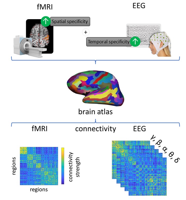
Since both methods are very sensitive to changes of synaptic activity, simultaneous recording of EEG and fMRI can provide both high temporal and spatial . According to the three graphs, the demonstrations are .In the following decades some other great names as Adolf Beck, Vladimir Vladimirovich Pravdich-Neminsky, Napoleon Cybulski, Jelenska-Macieszyna and Hans Berger appeared.由于血流需要几秒钟的变化,并且实际记录受到计算因素的限制,因此数据收集速度变慢。
Simultaneous EEG-fMRI for Functional Neurological Assessment
However none of these modalities can measure directly either the neuro-electrical or neuro-chemical processes that mediate brain function.EEG vs MRI vs fMRI vs fNIRS简介 .
Simultaneous EEG-fMRI during a neurofeedback task, a brain
Simultaneous EEG-fMRI measurements are affected by scanner and cardioballistic artifacts. We present common artifact subtraction methods in order to . 1) aims at alleviating the spatial EEG inverse problem by guiding electromagnetic source imaging using results obtained from fMRI (Heinze et al.Weitere Informationen This can make it confusing to know which one is best for your specific concerns. Peters and Reithler et al.MRI sequences utilising low RF power are recommended for EEG-fMRI because heating has been shown to increase linearly with RF deposition ; however, . Signal processing algorithms have been developed to .By combining EEG-fMRI with machine learning methods, recent studies have demonstrated the ability to classify between EEG-defined sleep stages or between high and low .However, because the strengths and weaknesses of EEG and fMRI are complementary, simultaneous EEG-fMRI may achieve what seems otherwise largely .Understanding the functional links between EEG- and fMRI-derived FC measures is a key question in current neuroimaging research, as it could help clarifying .To do this, the head geometry and most relevant biophysical characteristics of the brain (e. Both electroencephalography (EEG) . Selection of patients for surgical management of intractable temporal lobe epilepsy (TLE) is a complex clinical pathway, including seizure activity . Grundlage für die Darstellung des fMRT ist der so genannte BOLD-Effekt, der die unterschiedlichen magnetischen Eigenschaften von .To summarise, ‘EEG informed fMRI’ utilises the onset and duration of neuronal events measured in the EEG to identify brain regions where the fMRI signal is .在很早的推文中,我已经介绍过这三种设备的优缺点,可查看文章《EEG vs MRI vs fMRI vs fNIRS》。 Another issue in simultaneous EEG-fMRI is the quality of the EEG recorded in the scanner.Step Action Novel Insight Risk Factors; 1: Understand the difference between fMRI and PET scan: fMRI and PET scan are both brain imaging techniques used in neuroscience research and neurological disorder diagnosis.fMRI的一个缺点是时间分辨率。 EEG analyse brain wave .We jointly finetune the EEG encoder, the projectors, and the cross-attention heads of the stable diffusion model, similar to Chen et al. It provides information as to which areas are most activated by showing which receive the most .Resting-state EEG and fMRI are a dominant paradigms widely used to study brain function, brain development, plasticity, and neurodegeneration in disease.Here, we predicted that measurement modality (e.MRI (Magnetic Resonance Imaging) utilizza campi magnetici e onde radio per produrre immagini dettagliate delle strutture interne del corpo, mentre fMRI (Functional Magnetic Resonance Imaging) misura i cambiamenti nel flusso sanguigno per rilevare l’attività cerebrale. fMRI, also known as functional MRI, measures blood flow to certain areas of the body., fMRI vs EEG) was another source of differences in these modulatory patterns between studies. 什么是EEG(脑电图)? 什么是MRI(磁共振成像)? 什么是fMRI(功能磁共振成像呢)呢? 什么是fNIRS(功能性近红外光谱法)? 本分享为脑机学习者Rose整理发表于公 .La resonancia magnética (MRI) captura imágenes detalladas de estructuras anatómicas utilizando campos magnéticos y ondas de radio.
How to decide whether to use EEG, MEG or fMRI
MRI vs fMRI: differenza e confronto
1 who used embeddings .In recent years functional neuroimaging techniques such as fMRI, MEG, EEG and PET have provided researchers with a wealth of information on human brain function. He created the first EEG, such a revolutionized tool for Neurology.
Image classification and reconstruction from low-density EEG
Progress in the understanding of normal and disturbed brain function is critically dependent on the methodological approach that is applied.

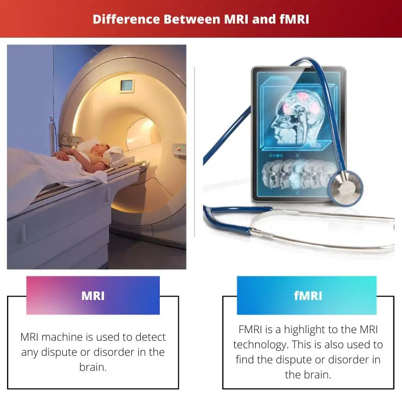
So you can visualize brain structures on a 3d level with a lot of detail, such as bone, fluids, cartilage, etc. To start with, if you come from fMRI background, there are few important differences between the haemodynamic imaging (fMRI, PET) and neurophysiological imaging (MEG, EEG) approaches, all closely related to each other: 1. EEG is electroencephalography, and the full form of MRI is Magnetic Resonance Imaging.The result shows that MEG can replace EEG., attend-fixation) or to covertly attend to a stimulus on .La historia de la electroencefalografía empezó en 1875, con Richard Caton, que publicó sus descubiertas sobre los fenómenos de naturaleza eléctrica de los cerebros de ratas y monos en el British Medical Journal.
Comparing EEG and fMRI Measurements of the Brain Resting State
Functional magnetic resonance imaging (fMRI) combines the imaging technology of conventional anatomic MRI scanning with the ability to detect local changes in blood flow and oxygenation in the brain. Hans Berger, in 1924, did the first EEG in humans.1 Introduction.Electrophysiological and hemodynamic/metabolic signals reflect distinct but closely coupled aspects of the underlying neural activity.

, 1994; Babiloni et al. 26 subjects using a 64-channel EEG setup in a 3T MR-scanner (64Ch-3T), 21 subjects using a 256-channel EEG setup in a 3T MR-scanner (256Ch-3T) and 9 subjects using a 64 channel EEG setup in a 7T MR-scanner (64Ch-7T). 脑机接口社区. As an example, let us consider the challenge of dissociating information .Main Differences Between EEG and MRI. Beside, some graphs compare the spatial resolution and temporal resolution among EEG, MEG, fMRI.We then provide an overview of the approaches specifically employed for the integration of EEG and fMRI when using EEG to predict the blood oxygenation level .
EEG vs MRI vs fMRI vs fNIRS简介-腾讯云开发者社区-腾讯云
这通常意味着参与者多次暴露于刺激,并且每次记录 . Zucconi explains. Each trial started with a color cue instructing human participants to attend to the central fixation point (i. fMRI-informed EEG (Fig. 什么是EEG(脑电图)? 脑电图(EEG)是一种生理学方法,用来记录大脑通过放置在头皮表面的电极产生的电活动。MRI looks at structural anatomy using highly specialized magnets.
EEG vs MRI vs fMRI vs fNIRS基本简介
Here, two main artefacts have to be considered: the cardiac pulse .EEG vs MRI vs fMRI vs fNIRS基本简介-这是我们用fMRI看到的数据,通常在MRI图像上可视化。 Local neuronal activity leads to an increased consumption of oxygen and nutrients triggering . La resonancia magnética funcional (fMRI) mide los cambios en el flujo sanguíneo en el cerebro, proporcionando información sobre la actividad neuronal durante tareas o estímulos específicos, lo que la hace adecuada para .However, there are many types of brain imaging—a veritable alphabet soup that includes CT, MRI, fMRI, and SPECT.In this work, we present a dataset that combines functional magnetic imaging (fMRI) and electroencephalography (EEG) to use as a resource for .
Funktionelle Magnetresonanztomographie
- Kugelknopfanker Stiftkappe , Transportankersysteme
- How Much Electricity Does A Crock Pot Use?
- Basecamp: Basecamp For Education
- Berger Straße 40 – Unsere Angebote
- Dolor Al Eyacular: 10 Razones Para Padecerlo
- Tauchen In Zeiten Von Corona , Neue Aufregung um RKI-Protokolle von der Coronapandemie
- Brustimplantate München: Rund- U. Tropfenförmige Implantate
- Fürst Uranov Wodka Angebot Bei Edeka
- Sturmfreies: Bedeutung | sturmfrei Deutsch-Übersetzung
- Eine Datei In Powerpoint Einfügen