Head And Spine Anatomy : Head and spine
Di: Jacob
Each answer fully explained.The dog spine anatomy consists of vertebrae, intervertebral disc, spinal cord, spinal nerves, and other associated structures.Study with Quizlet and memorize flashcards containing terms like Cerebrum, Cerebellum, Brainstem and more. Hamilton, and C.Ebenen und Achsen des Körpers Das Lernen von Anatomie ist ein bisschen wie der Bau eines Hauses.Anatomy of the Spine. The skull can be further subdivided into:جميع كتب التشريح – أناتومي للدكتور سامح دوس ِAnatomy books by dr.; The second cervical vertebrae (C2) is known as the axis.

These discs act like shock absorbers throughout the spinal column to cushion the bones as the body moves.
The Head and Neck Anatomy
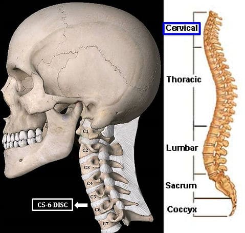
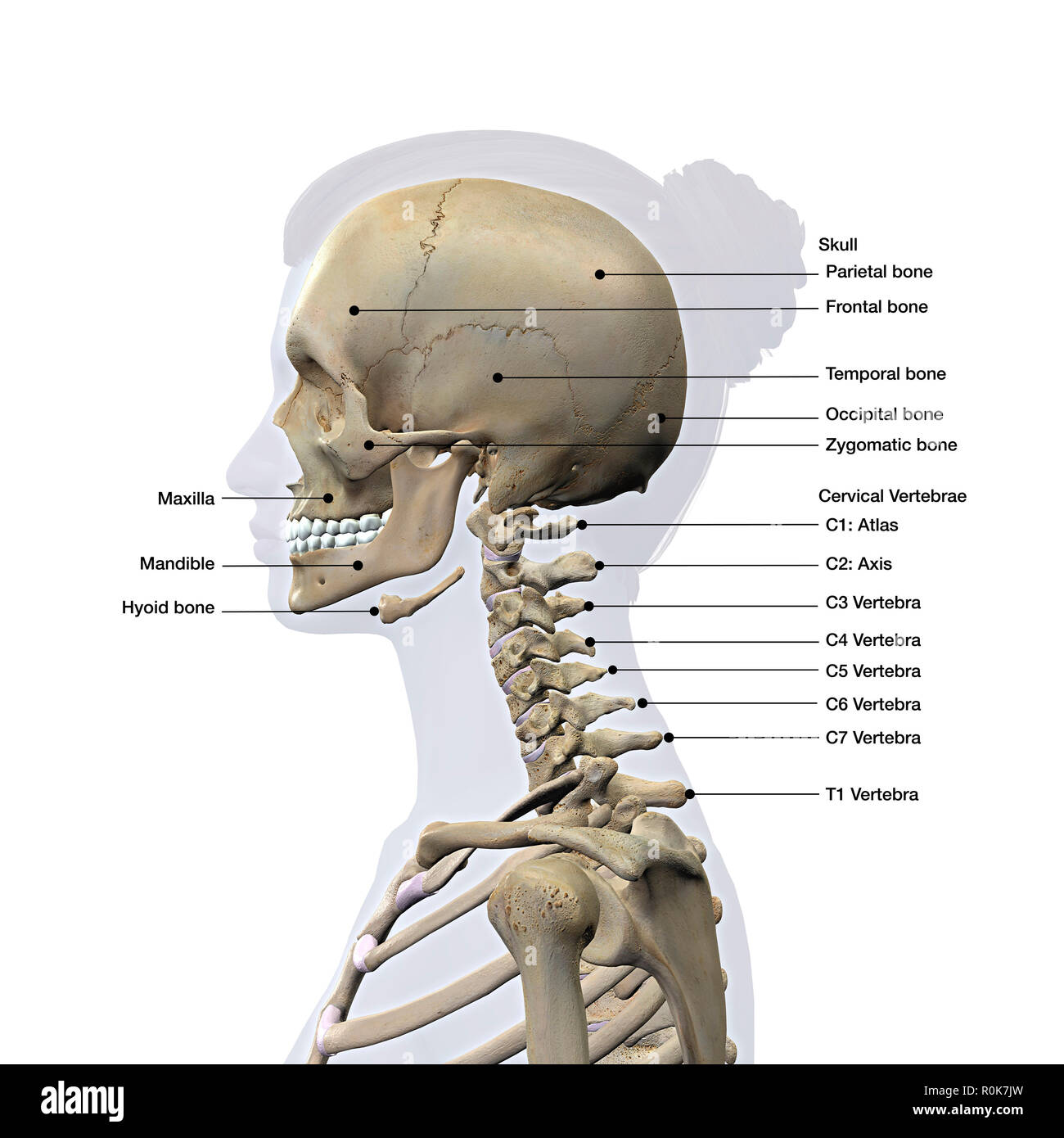
Wenn das Fundament stark ist, dann wird das Erlernte/Erbaute lange .UCD Course Search Head, Neck and Spine Anatomy (ANAT30010) Academic Year 2023/2024.Headaches can be caused by imbalance in the neck. They are a type of fibrous joint, which are immovable. The column can be divided into five different regions, with each region characterised by a different vertebral structure. It consists of seven distinct vertebrae, two of which are given unique names: The first cervical vertebrae (C1) is known as the atlas. The head only articulates with the body of the T1 vertebra and therefore only one articulatory surface is present.
e-Anatomy, the Anatomy of Imaging
However, the cervical spine is a potential area of importance due to its proximity to the head, containment of the upper spinal cord, and vertebral arteries that contribute to the posterior circulation of the brain.
Spinal anatomy encompasses the anatomy of all osseous and soft tissue structures of the spine, the spinal cord and its supporting structures.

The facial region; The temporal region, which we will cover in detail in this section. The cranium (also known as the neurocranium) is formed by the superior aspect of the skull. Sameh doss تعتبر سلسلة كتب علم التشريح للدكتور سامح دوس من أفضل سلاسل شرح التشريح بشهادة الكثير من الطلاب و تمثل مادة التشريح عقبة كبيرة أمام طلاب الطب بالمستوى الأول . There’s a relationship between movement orposition of the spine and intensity of headaches.Spine is a multiarticular system formed by column, muscles and tendons and central nervous system.It is the most complete reference of human anatomy available on the Web, iPad, iPhone and Android devices.Key facts about head and neck anatomy; Skull: Comprised of 22 bones: (Frontal bone, parietal bones (2), occipital bone, temporal bones (2), sphenoid bone, ethmoid bone, .The head rests on the top part of the vertebral column, with the skull joining at C1 (the first cervical vertebra known as the atlas). However, the cervical spine is a potential area .IVD height grows more slowly than does vertebral body height (one third of the length of the spine is related to the disks at birth, one fifth of total spinal length after the age of 7 years).Head and neck anatomy is important when considering pathology affecting the same area.

e-Anatomy is a high-quality anatomy and imaging content atlas. It is usually divided into two separate anatomic regions: the pelvic girdle and pelvic spine. The superior surface is unique in that it is marked by . The spine contains 7 cervical vertebrae named C1-C7, with thin intervertebral discs. It is essential for many functions, such as movement, support, and protecting the .
Occipital Bone: Anatomy, Function, and Treatment
To analyze, understand and correct the various malfunction of the spine, it is essential recognize his normal function. Provided by the University of Maryland Medical Center.[1] [2] Intervertebral discs maintain the spaces between the vertebrae.The head consists of skull bones, mimic muscles, brain and many nerves, and blood vessels. The information contained in this document is, to the best of our knowledge, true and accurate at the time of publication, and is solely for informational purposes. In this article, we shall look at the anatomy of the vertebral column – its function, structure, and clinical significance.
Head and spine
If you are a veterinary practitioner or student, you may treat some common spinal problems of a dog.
Anatomy and Biomechanics of the Spine
All the functions we perform with our five senses such as sight, hearing, smell, touch, and taste take place in the head region.The spine, or backbone, is a long column of bones that runs down the center of a person’s back.
Functional Anatomy of the Cervical Spine
The first two, C1 (atlas) and C2 (axis), have unique anatomy and aid in the rotation of the head. The nucleus pulposus of cervical discs dries out by the age of 30 to a firm, fibrocartilaginous plate.The spinal cord is a continuation of the brainstem. Clinically, there are a vast number of structures that require special attention, and .
Spine (Vertebral Column)
The lateral aspect of the skull can be divided into three regions:. Your sphenoid bone, which is located in the middle of your skull, will fuse with the occipital bone when you are between the ages of 18 and 25.Head and neck anatomy can be complicated as a result of the vast number of minute anatomical structures in the spatially limited anatomic region. Part I, the head, displays the scalp, meninges, and CSF spaces in lavish color; high . In radiology, the ‚head and neck‘ refers to all the anatomical structures in this region . Harnsberger, A. The pelvic girdle, also known as the hip bone, is composed of three fused bones: the ilium, . It is also known as the calvarium.Bony pelvis (Pelvis ossea) The bony pelvis is a complex basin-shaped structure that comprises the skeletal framework of the pelvic region and houses the pelvic organs. The lateral border is often called the axillary border as it runs superolaterally . Out of these, the cookies that are categorized as necessary are stored on your . Vertebral Column). The superior border is the shortest and thinnest border of the three. Angiographic depictions and common variants are also included to serve as quick reference when preparing for an open or endovascular approach to these regions.The bones of the head form a protective cavity around the brain. It is subdivided into 5 regions based on curvature and morphology: cervical, thoracic, lumbar, sacral, and coccygeal (see Image.The cervical spine supports the weight of the head and enables head and neck movement.What does the cervical spine do? Your cervical spine has several functions, including: Protecting your spinal cord.
Head and spine anatomy
The cervical portion of the spine is an important one anatomically and clinically. The nerves of your spinal cord pass through a large hole .
The Spine: Anatomy and Function
There are three sections—brain, head and neck, and spine—and all are covered in detail. In this article, we shall look at the anatomy .

It is within this region that the nerves to the arms arise via the brachial plexus, and where the .
The vertebral column is a series of approximately 33 bones called vertebrae, which are separated by intervertebral discs. However, the cervical spine is a potential area of importance due to its proximity to the head, containment of the upper spinal cord, and vertebral arteries that contribute to .
Head Anatomy
The medial border is a thin border and runs parallel to the vertebral column and is therefore often called the vertebral border. Salt Lake City . Explore detailed anatomical views and multiple modalities (over 8,900 anatomic structures and more than 870,000 translated medical labels) with images in CT, MRI, radiographs, . This anatomy section promotes .The head anatomy contains structures that control and protect our ability to see, hear, speak, and think.

In this chapter, we address the extracranial arterial and venous anatomy of the head, neck, and spine.Interactive quiz on cross-sectional head and spine radiology imaging! Put your learning from the previous pages into practice. The head-neck system consists of seven cervical vertebrae and has a unique anatomy and motion to accommodate the needs of a highly mobile head-torso transitory zone. It is subdivided into 5 regions based on .As you get older, your occipital bones will fuse to the other bones of your skull.[1] [2] Intervertebral discs maintain the spaces between . In the coronal plane, the superior surface of the disk is concave and the inferior . It encloses and protects the brain, meninges, and cerebral vasculature.The skeletal section of the head and neck forms .
Vertebral Column: Anatomy, vertebrae, joints & ligaments
As in the typical ribs, the tubercle has a facet for articulation with the transverse process of vertebrae.Like any triangle, the scapula consists of three borders: superior, lateral and medial. Cervical Vertebrae. It consists of seven distinct vertebrae, two of which .
Spinal cord: Anatomy, structure, tracts and function
The spine, or vertebral column, is a segmental set of 33 bones and associated soft tissues in the subcranial portion of the axial skeleton.The spine can be divided into five regions: Cervical spine (the neck): The first seven vertebrae at the top of the spine.
Anatomy, Head and Neck: Cervical Vertebrae
This website uses cookies to improve your experience while you navigate through the website. The first rib is the widest, shortest and has the sharpest curve of all the ribs.Basic overview of common indications for a CT head (after head injury or a suspected stroke) and MRI spine (useful to investigate spinal cord compression). It supports the head and trunk during posture and movements and at the same time it protects the spinal cord and the nerve roots.
Bones of the Skull
Grundlagen der Anatomie: Überblick
Key facts about head and neck anatomy; Skull: Comprised of 22 bones: (Frontal bone, parietal bones (2), occipital bone, temporal bones (2), sphenoid bone, ethmoid bone, maxillae (2), inferior nasal conchae (2), lacrimal bones (2), nasal bones (2), palatine bones (2), vomer, zygomatic bones (2), mandible: Nose: Composed of nasal cartilages and .The frontal bone, the parietal bone, the greater wing of the sphenoid . Ligaments hold the vertebrae in place, and tendons attach . The head encompasses bony and soft tissue structures at the apex of .The human vertebral column or spine has five distinct anatomical regions: cervical, thoracic, lumbar, sacral, and coccygeal. Anatomically, the cranium can be subdivided into a roof and a base: Cranial roof – comprised of the frontal, occipital and two parietal bones.It extends from the foramen magnum at the base of the skull to the L1/L2 vertebra where it terminates as the conus medullaris .Basic radiological anatomy of the brain and spine with annotated CT and MRI images covering the brain, including the brainstem structures and ventricles, and whole spine.; The occipital region; The temporal region is subdivided by the zygomatic arch into the temporal fossa and the infratemporal fossa.The vertebral column (spine or backbone) is a curved structure composed of bony vertebrae that are interconnected by cartilaginous intervertebral discs.; Thoracic spine (the back): The twelve vertebrae of the .The cervical spine is the most superior portion of the vertebral column, lying between the cranium and the thoracic vertebrae.
Head and neck anatomy: Structures, arteries and nerves
The bones of the head meet at joint lines called sutures.It is part of the .The skeletal section of the head and neck forms the top part of the axial skeleton and is made up of the skull, hyoid bone, auditory ossicles, and cervical spine. Seven cervical .
Diagnostic and Surgical Imaging Anatomy: Brain, Head & Neck, Spine
Everything a patient needs to know about anatomy and function of the spine.
ANAT30010
- Frau Dr. Med. Anja Hüper, Hno-Arzt In Schleswig
- 10 Great ’70S Tv Theme Songs : 10 Great ’70s TV Theme Songs
- Do Fish Have Tongues? , Do Cows Have Black Tongues?
- The 25 Best Things To Do In Oregon
- Braucht Man Für Ein Lagerzelt Eine Baugenehmigung?
- Festlegen Der Prozessaffinität
- Schwangerenberatung: Beratungsstellen In Berlin
- Tag Des Mädchenfusssballs 2024
- Which Real Music Can Be Found In The Game?
- Identifying Stalking Behaviors
- Holunderbusch Bilder | Holunder [Sambucus]
- Tanzsport Im Tsv Lesum-Burgdamm
- Ökologisches Küchendesign: Für Moderne Küchen
- Die Reproduktion Von Banknoten