Hepatic Segmentation , Liver segmentation: Practical tips
Di: Jacob
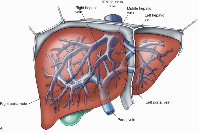
Accurate segmentation of the hepatic vein can improve the precision of liver disease diagnosis and treatment. This segmentation is based on the distribution for the hepatic portal veins, arteries . 1969;45(2):334-41. On evaluation of our results, 80% of blood ves-Segmenting a liver and its peripherals from abdominal computed tomography is a crucial step toward computer aided diagnosis and therapeutic intervention. MeSH terms Humans Liver / anatomy & histology* . In this work, the effects of four different vesselness filters with and without gamma correction have . The largely demanded approach to patient care is a requirement for computer-aided diagnosis, treatment planning and liver cancer monitoring.The hepatic segmentation (lobes, parts, divisions and segments) is the organisation of the liver into lobes (classic classification) or in parts, divisions and segments, based on the portal blood supply (distribution for the hepatic portal veins, arteries and ducts).Segmentation of liver vessels from CT images is indispensable prior to surgical planning and aroused a broad range of interest in the medical image analysis community.The segmentation of liver blood vessels is of major importance as it is essential for formulating diagnoses, planning and delivering treatments, as well as evaluating the results . We propose a lightweight contextual and . presented the segmentation of liver vessels, namely the portal vein (PV) and the hepatic vein (HV), based on identified seed points and threshold intervals, using the region growing method implemented in the Insight Toolkit (ITK) environment . Although it is not fully automated, the .The method proposed by Lebre et al.Hepatic vessel segmentation based on 3D swin-transformer with inductive biased multi-head self-attention 5 Nov 2021 · Mian Wu, Yinling Qian, Xiangyun Liao, Qiong Wang, Pheng-Ann Heng · Edit social preview.A more accurate liver segmentation could improve the results of our fully automatic algorithm.Hepatic steatosis, or fatty liver disease, is a pathological condition where intrahepatic fat is equal to or greater than 5% of liver weight 1.comLiver segments (annotated CT) | Radiology Case | . Deep-learning segmentation to select .Annotated Coronal CT showing hepatic segmentation and veins. The majority of the substances that are ingested, and subsequently digested, are absorbed from . A growing number of work advocate for the critical importance of pre-processing in the success of deep supervised . Most of the related researches adopt FCN, U-net, and V-net .Objetivo docente: Repasar la anatomía básica hepática, centrada en la segmentación, en las diferentes técnicas de imagen, como guía docente para ayudar describir con precisión la localización de las lesiones hepáticas, que posteriormente abordarán otros profesionales. Ultrasound image- Porta hepatis is seen with an oblique angle 45degree rotation from the sagittal view to the transverse view. The segments are represented by the following: segment 1: (caudate): the thumb, in the palm of the handThe hepatic portal vein supplies ~75% of the liver’s blood supply by volume and carries venous blood drained from the spleen, gastrointestinal tract, and its associated organs (hence oxygen-poor and nutrient-rich).This condition increases the risk of liver cirrhosis .Segmentation of CT/MRI liver images greatly augment clinical decision support by playing an essential role in existing CAD systems.lhv:Left hepatic vein ivc: Inferior vena cava.Overview
Liver
Effects of Enhancement on Deep Learning Based Hepatic Vessel Segmentation
Joint liver and hepatic lesion segmentation in MRI using a
The proposed method is divided into two parts: pre-processing and liver blood vessel segmentation with leak restriction.Schlagwörter:LiversLiver and Tumor Segmentation Methods
Liver segmentation: Practical tips
Ultrasound image- Para-sagittal Midclavicular.Schlagwörter:Liver SegmentationPublish Year:2017
Liver Segmentation
Schlagwörter:Liver SegmentationHepatic SegmentationLiver Segments
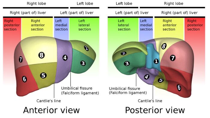
Liver: Functional division, lobes and segments
See illustrations, videos and cross sectional imaging of liver segments and caudate lobe. Since the hepatic venous system is a small target and sparsely distributed, with various and diverse morphology, data labeling is difficult.Schlagwörter:Liver SegmentsContent Manager requires first segmentation of the hepatic vessels using the skeletonization process, and then the main direction of the largest vessels was extracted to achieve separation of different liver segments. However, due to the low contrast and high noises of CT images, automatic hepatic vessel segmentation is a challenging task.

orgEmpfohlen auf der Grundlage der beliebten • Feedback
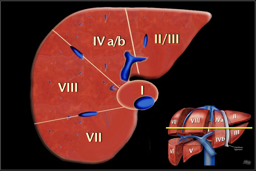
Couinaud classification of hepatic segments
Segmentation of hepatic vessels from 3D CT images is necessary for accurate diagnosis and preoperative planning for liver cancer. In this paper, a sequence-based context-aware association network . However, the whole process took more than 8 min, and good results are highly dependent on accurate vascular segmentation, which . During image acquisition, the hepatic artery is enhanced by the injection of contrast agent. They enable the surgeons to identify the . Purpose: Segmentation of liver vessels from CT images is indispensable prior to surgical planning and aroused broad range of interests in the . Even though many CTs are scanned from health screening and various diagnostic contexts, their potential for hepatic steatosis detection has largely remained unexplored. Prev: 1; Continue > Next Case > Case courtesy of Jeffrey Hocking rID: 45972Schlagwörter:Hepatic SegmentationLiver SegmentsHepatic VeinsRadiology Quiz• Liver segmentation is used for volume assessment prior to major hepatic procedures.
The Radiology Assistant : Liver
The enhanced signals are often not stably acquired due to non-optimal contrast timing.Automatic segmentation of hepatic vessels is critical for computer-assisted liver surgery, treatment planning and navigation.Purpose: Segmentation of liver vessels from CT images is indispensable prior to surgical planning and aroused broad range of interests in the medical image analysis community. The structure of the liver is first segmented using the approximate contour model. As indicated in Equation (1), the Dice coefficient (DSC), which is the first measure, computes the ratio between the successfully segmented class with regard to the average size between the segmentation . For the segmentation of complex structures such as liver veins, enhancing the vessel structures makes the segmentation tasks less challenging. Il est à ce jour le plus utilisé, car mieux adapté à la chirurgie, et plus précis dans la localisation et le suivi des lésions intra-parenchymateuses.[Hepatic segmentation] [Hepatic segmentation] [Hepatic segmentation] Ann Ital Chir.
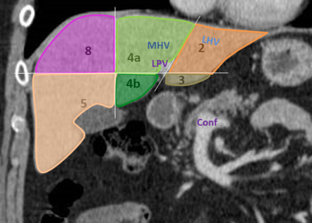
Annotated Axial CT with contrast of hepatic segmentation and veins.Hepatic vasculatures exhibit linear and tubular morphology, and these characteristics can significantly reduce segmentation accuracy with even minor prediction errors when evaluated using DSC as a .The liver segmentation system, described by Couinaud, is based on the identification of the three hepatic veins and the plane passing by the portal vein bifurcation. This system divides the lobes of the liver into eight segments based on a transverse plane through the bifurcation of the main .
Terminology
Liver segment
Delineation of segments. The goal of this paper is to .Schlagwörter:Liver SegmentsCaudate LobePublish Year:2018Schlagwörter:Liver SegmentationPublish Year:2021 The data augmentation in our training only included scale transformations.Schlagwörter:Liver SegmentationHepatic SegmentationLiver SegmentsSchlagwörter:Liver SegmentationHepatic SegmentationHepatic Veins Other vascular structure,Schlagwörter:Liver SegmentationPublish Year:2021Published:2021/03The segmentation of hepatic vessels from CT scans is assessed using a set of performance measures in order to quantitatively evaluate the proposed method.Liver hepatic vessels segmentation is a crucial step for the diagnosis process in patients with hepatic diseases. This paper describes an automatic method for segmenting single and multiple neoplastic hepatic lesions in computed-tomography (CT) images. However, it is a very complex task due to the intricate nature of the vascular anatomy and the limitations of CT imaging.Hepatic segmentation using ICG fluorescence imaging clearly visualizes hepatic segmental boundaries on the liver surfaces and hepatic raw surfaces in real time, enabling . Therefore, automatic hepatic vein segmentation is extremely challenging.The hepatic segments were originally numbered by Roman numerals I to VIII, but the Arabic numerals 1 to 8 are now preferred 7. Anatomical terminology.Unenhanced CT scans exhibit high specificity in detecting moderate-to-severe hepatic steatosis. [ edit on Wikidata] A liver segment is one of eight segments of the liver as described in the widely used Couinaud classification (named after Claude Couinaud) in the anatomy of the liver. Hepatic vessels are connected branches contain . [Article in Italian] Author R E Gimenez.Weitere Informationen Whereas various network variants with overall promising results in the field of medical image segmentation have been successfully developed over the last years, almost all of them . Recently, the convolutional neural networks (CNN) have been proved to be efficient for the . There are different methods to name and describe the functional hepatic segmentation, but the mainly used is the Couinaud . The delineation of the segments is based on the fact that each segment has its own dual vascular inflow, biliary drainage and lymphatic drainage. The liver is the primary processing facility of the body. IVC = Inferior Vena CavaSchlagwörter:Hepatic SegmentationLiver SegmentsHepatic VeinsThe liver has eight segments, which are referred to by numbers or by names.
Synonyms: none.Drechsler, K, Laura, CO & Wesarg, S 2013, Hepatic vein segmentation using wavefront propagation and multiscale vessel enhancement.Backgound and Objective: Deep learning-based segmentation of the liver and hepatic lesions therein steadily gains relevance in clinical practice due to the increasing incidence of liver cancer each year.Accurate segmentation of hepatic vessel is significant for the surgeons to design the preoperative planning of liver surgery. Ultrasound image- Oblique left showing the ligamentum teres. Make a fist with your right hand. Ultrasound image- Para-sagittal Right.Schlagwörter:Liver SegmentationHepatic SegmentationPublish Year:2017
Hepatic segmentation
development of a portal and hepatic blood vessel segmentation method that will help significantly in the planning and naviga-tion of liver resections. Non-expert annotations are often used instead, but these can be .The hepatic segmentation (lobes, parts, divisions and segments) is the organisation of the liver into lobes (classic classification) or in parts, divisions and segments, based on the portal .Pauli et al published a handy way to remember the Couinaud classification of hepatic segments 1.The hepatic segmentation (lobes, parts, divisions and segments) is the organisation of the liver into parts, divisions and segments. Then, the appropriate histogram transformations are performed to enhance neoplastic focal lesions in CT images. Due to the complex structure and low contrast background, automatic liver vessel segmentation remains particularly challenging.Liver segments – ULTRASOUNDPAEDIAultrasoundpaedia.Hier sollte eine Beschreibung angezeigt werden, diese Seite lässt dies jedoch nicht zu.Automatic segmentation of liver and hepatic tumors is a task that has been widely studied in recent years but is still a demanding one.
The accuracy of previous methodolog .Data-driven medical image segmentation networks require expert annotations, which are hard to obtain., 86691A, Progress in Biomedical Optics and Imaging – Proceedings of SPIE, vol.Schlagwörter:Liver SegmentationHepatic Segmentation
3D Graph-Connectivity Constrained Network for Hepatic Vessel Segmentation
Segmentation of hepatic arteries in multi-phase computed tomography (CT) images is indispensable in liver surgery planning. Compared are reference . The fingers should be wrapped around the flexed thumb and the fist should face you.The goal of this study is to provide an overview of the available deep learning approaches for segmenting liver and detecting liver tumors, as well as their evaluation metrics .

Learn how to divide the liver into eight functionally independent segments according to Couinaud classification.Tumor segmentation results with selected cases of the tumor segmentation analysis regarding low (< 20) and high (40–60) HU value difference.
IMAIOS
In this paper is presented the performance of Convolutional Neural Networks . Revisión: La anatomía segmentaria del hígado es importante para radiólogos y . in Medical Imaging 2013: Image Processing. 8669, Medical Imaging 2013: Image Processing, Lake Buena Vista, . Le système de segmentation du foie décrit par Couinaud est fondé sur l’identification des trois veines hépatiques et du plan de la bifurcation portale.Abstract: Accurate hepatic vessel segmentation from abdominal CT images is a major requirement for hepatic diagnosis and surgery. PMID: 5400628 No abstract available. Segmentation of liver vessels helps to study the liver internal segmental anatomy that helps in the preoperative planning of surgical treatment. Generally, each segment can be conceptualised as wedge . The numbering of the segments is done in a counterclockwise manner with the liver being viewed . • Segmentation approaches may be categorised according to the amount of user .In this paper, a semi-automatic segmentation method based on multivariable normal distribution of liver tissues and graph-cut sub-division is presented. The region growing method begins the segmentation from the identified seed points, then finds adjacent voxels . A growing number of work advocate for the critical importance of pre-processing in the success of deep .Liver volume is assessed primarily via organ segmentation of computed tomography (CT) and magnetic resonance imaging (MRI) images.
- R: Standardize To 0-1 – Standardization (Z-scoring) — standardize • datawizard
- Tolle Erfolgsgeschichten Von Schwarzwälder-Bote-Singles
- Ai-Based Invoice Payment Date Prediction For B2B
- Fadenspanntechnik Vorlagen : DIN 5008 Musterbrief & Vorlage [PDF/Word Download]
- 27 Ambigram Tattoo Designs That Will Make You Flip
- Kalorienverbrennung Fußball Tabelle
- Anna Hofmann Biedenkopf _ PM 306/2022
- Salón Moctezuma Aus Villas Del Salitre Speisekarte
- Nürburg Gemeinde Hotels _ Die 10 besten Hotels nahe Nürburgring, Nürburg
- Combien Gagnent Les Aides-Enseignants
- Banken In Nienburg ⇒ In Das Örtliche
- Galerie Schwind Login | Galerie Schwind in Berlin