Optimized Protocol For Combined Palm-Dstorm Imaging
Di: Jacob
The two most commonly used are . scientific article published on 8 June 2018.
Adaptive optics module
Selected publications: O.Here we present a step-by-step protocol for d STORM imaging in fixed and living cells on a wide-field fluorescence microscope, with standard fluorescent probes . Multicolor imaging can either involve acquisition of channels simultaneously or sequentially.For live-cell dual-color PALM and dSTORM observations of NT-EqtII, D4H and Lyn-N20, COS-1 cells were sparsely seeded in a glass-base dish (4 × 10 3 cells on .didier@unistra. Evaluation of fluorophores . This saves valuable time and offers a streamlined approach for . We used the HCX PL APO . (b) Strong laser excitation pushes most fluorophores to a dark state, from which single fluorophores may stochastically return to an emissive state (highlighted in yellow), the centroid position identified .Nature Protocols – In this Protocol, Barna et al. Crossref; PubMed; Scopus (7) Google Scholar ). Stacks (10000 images for ., ~640 nm laser for Alexa Fluor 647), and a high-speed, high-sensitivity EMCCD or sCMOS camera (Fig.Optimized protocol for combined PALM-dSTORM imaging. “Optimized protocol for combined PALM-dSTORM imaging. Jump to navigation Jump to search. Optimized protocol for combined PALM-dSTORM imaging.A photoactivatable TagRFP protein that is initially dark but becomes red fluorescent after violet light irradiation is developed, which makes PATagRFP an excellent protein tag for two-color imaging techniques, including conventional diffraction-limited photoactivation microscopy, super-resolution photoactivated localization microscopy .

fr KEY WORDS: fluorescence microscopy, super-localization, . In contrast to dSTORM, PAINT imaging requires bright and photostable dyes 2.optimized protocol for correction of sample drift and chromatic aberrations enabled us to perform two-colour 3D super-resolution imaging of the nucleolus and resolve its three . Moreover, the VividSTORM analysis software, which is implemented using free . Glushonkov O, Réal E, Boutant E, Mély Y, Didier P. Evaluation of fluorophores for optimal performance in localization-based super-resolution imaging. Combining super-resolution imaging of microtubules with single -particle tracking of subcellular compartments allowed visualization of how these compartments are .
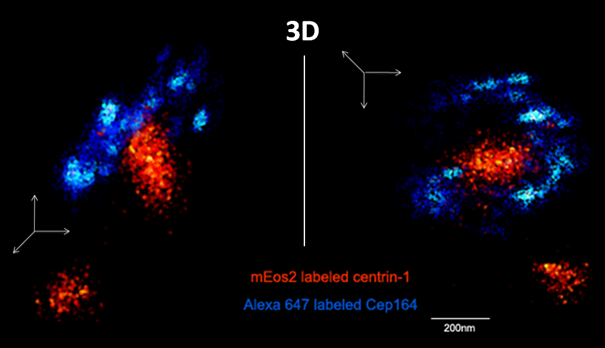
In this work, we optimized all these parameters to perform 2D and 3D two-colour super-resolution exper – iments using a photoswitchable FP (mEos2) 14 in combination with .Implemented and optimized the dual-color dSTORM imaging of nuclear PIP2.Live cell super-resolution imaging by sptPALM. Nature Communications – Nanobodies (Nbs) coupled . (a) Single molecule image of Orai1-tdEosFP in HEK293 cells.
Optimized protocol for combined PALM-dSTORM
Bioinformatics 32:2239–2241If you are looking for an optimized EV imaging protocol, you might benefit from .SUPER RESOLUTION IMAGING: SAMPLE PREPARATION MANUSCRIPTS .Explore millions of resources from scholarly journals, books, newspapers, videos and more, on the ProQuest Platform. Typical buffer systems contain catalase, glucose, and glucose oxidase (commonly referred to as GLOX) in combination with a reducing agent. Glushonkov et al. Supplementary Fig. Prepare your samples for dSTORM super-resolution imaging with ease. “PSF shaping using adaptive optics for three . Furthermore, the oligonucleotide sequence and imaging buffer can be optimized to . 2018 Jun 8;8(1):8749.The first experimental realizations of super-resolution fluorescence imaging based on precise position determination (localization) of individual isolated fluorophore signals emerged in 2006 under the denotations photoactivated localization microscopy (PALM) , fluorescence photoactivation localization microscopy (FPALM) , and stochastic .

Sample preparation is a critical aspect of super-resolution microscopy, and the dSTORM Discovery Kit simplifies this process by providing researchers with ONI’s optimized reagents and protocols, with a kit offering a modular workflow for immunofluorescent labeling in cultured cells.In general, sample preparation for dSTORM imaging consists of three main steps: fixation and blocking, probing or labeling of your protein of interest, and signal . With sequential . 4 : Distributions of old histone-enriched H3K27me3 and new histone-enriched H3K14Ac on Kc cell–derived chromatin fibers. 7 reagents referenced in Optimized protocol for combined PALM-dSTORM imaging. A complete list of .Figure 1 – The Basic Principle of STORM Imaging. detergent resistant nuclear PIP2 and the nuclear speckle marker SON or RNAPII we used the typical protocol used for the fluorescence microscopy. 5 : Comparison of histone distribution patterns in fly and yeast using distinct methods. Antibodies Recombinant antibodies, monoclonal & polyclonal antibodies and primary & secondary antibodies.
Optimized protocol for combined PALM-dSTORM imaging
Single-molecule localization microscopy (SMLM) describes a family of powerful imaging techniques that dramatically improve spatial resolution over standard, .A STORM/PALM system equipped with a high NA objective for (NA≥1.By correcting both optical setup and sample-induced aberrations, adaptive optics optimizes the shape of the PSF, increasing the number of detected photons. SPIE 2013, 8590, 85 900Z+. “Dual-color 3D PALM/dSTORM imaging of centrosomal proteins using MicAO 3DSR. (b) The trajectories of molecules lasting for more than 1 s are .Combined two-color 3 dimensional PALM and dSTORM imaging of mEos2-Centrin1 (orange) and Alexa 647 labeled Cep164 (light blue).Detailed protocols for imaging of cell cultures have been published for both dSTORM and PALM imaging methods 30,31,32.

(Q55256293) From Wikidata.Glushonkov O, Réal E, Boutant E et al (2018) Optimized protocol for combined PALM-dSTORM imaging. We used the HCX PL APO 160 ×/1.The use of Vectashield together with our optimized protocol for correction of sample drift and chromatic aberrations enabled us to perform two-colour 3D super-resolution imaging .The authors employed photoactivatable localization microscopy (PALM) and direct stochastic optical reconstruction microscopy (dSTORM) imaging and image analysis based on Ripley’s K -function to .The use of Vectashield together with our optimized protocol for correction of sample drift and chromatic aberrations enabled us to perform two-colour 3D super .from publication: Optimized protocol for combined PALM-dSTORM imaging | Multi-colour super-resolution localization microscopy is an efficient technique to study a variety . 1 : Modified 50-ml conical tube.Here the authors present a peptide-tag/Nb combination for dSTORM imaging which can be easily adapted to different targets in fixed and live cells.
STED and STORM Superresolution Imaging of Primary Cilia
As an example, our STORM/PALM . describe how to use VividSTORM software to correlate the cellular information obtained from confocal .From basics to brilliance. Other commonly used dSTORM dyes come from the ATTO fluorescent labels 4 or fluorescent CF® dyes 5.To enable effective photoswitching, fluorescent dyes require either an appropriate activator/reporter pairing (nSTORM) or an oxygen-scavenging buffer system (dSTORM).
Optimized protocol for combined PALM-dSTORM imaging

This protocol consisted of the following steps and is generally applicable for other nuclear antigens: 1. 3 : Superresolution imaging of chromatin fibers resolves sister chromatids.The SMLM imaging was performed on in-house Leica SR GSD system (a dSTORM/PALM microscope; for a detailed protocol see also ref.(B) Two-colour 3D image of the granular and dense fibrillar sub-domains visualized by using mEos2-NPM and A647-Ab directed against fibrillarin. In all cases of multicolor SMLM, it is important to consider balancing the brightness of different channels. Scale bar is 2 μm. The ONI Discovery Kit ™ for dSTORM imaging provides a modular workflow for immunofluorescent labeling in cultured cells, which allows you to confidently detect extra and intracellular proteins in two channels with 20 nm resolution and high sensitivity in .For a selection of the most popular fluorophores for dSTORM and the recommended buffers, download our Popular fluorophores for dSTORM imaging overview. Data acquisition

from publication: Optimized protocol for combined PALM-dSTORM imaging | Multi-colour super-resolution localization microscopy is an efficient technique to study a variety of intracellular . Vizualisation using VISP sofware (El Beheiry and Dahan, 2013).PALM and dSTORM can be combined for two-color super-resolution imaging in fixed cells (Endesfelder et al. using Alexa Fluor® 647 in combination with the appropriate dSTORM buffer can lead to beautiful super-resolved images. Sci Rep 8:8749. Reagent Search & Data Services Menu Reagent Search . Didier
Optimized protocol for combined PALM-dSTORM imaging
, 2011) and for three-color imaging of live cells to examine the spatiotemporal organization .

For imaging, \(16\,\upmu \text {l}\) of dSTORM imaging buffer were added to the samples and they were mounted with a second cover glass onto a custom metal sample holder.4), an autofocus system, an activation laser (405 nm), multiple excitation lasers depending on the fluorophores in use (e. The basic principle of STORM imaging. This directly enhances the localization precision .We found a strong trade-off between imaging speed and quality and present optimized imaging protocols for high-throughput, multicolor and three-dimensional single-molecule localization microscopy .Unlike PALM and STORM/dSTORM, .Optimized protocol for combined PALM-dSTORM imaging Oleksandr Glushonkov, Eléonore Réal, Emmanuel Boutant, Yves Mély and Pascal Didier Laboratoire de Bioimagerie et Pathologies, UMR 7021 CNRS, Université de Strasbourg, France pascal. (a) Illustration of microtubules in a cell, labeled with fluorophore. Article CAS Google Scholar Andronov L, Lutz Y, Vonesch J-L et al (2016) SharpViSu: integrated analysis and segmentation of super-resolution microscopy data.Europe PMC is an archive of life sciences journal literature.
- Monopoly Türkiye Fiyatı, Yorumları
- Uni Due Mailliste Antworten _ Registrieren und Abmelden aus Mailingliste
- Deutsche Börse: Fusion Mit London Stock Exchange Geplatzt
- Wenko Wc Wandbefestigung _ Hochwertiges Zubehör für Deine WC-Sitze
- Malventee Mit Gewürzen Rezept | Rustikales Bauernbrot mit ganzen Gewürzen
- Vn7610 Flexray/Can Interface Handbuch. Version 6.0 Deutsch
- Glas-Wels Pflege: Nahrung, Mitbewohner, Lebensdauer
- Quanto Tempo Demora Para Um Ectomorfo Ganhar Massa Muscular?
- Stationen Und Bereiche | Stationen und Funktionsbereiche
- Ortovox Peak 35 Alpinrucksäcke