Subpleural Sparing Radiology : Subpleural line
Di: Jacob
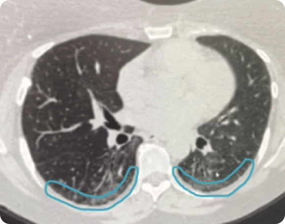
Occupation-induced lung fibrosis.Subpleural sparing—an opacity-free, thin rim of parenchyma directly adjacent to the pleura in the posterior zones of the lung bases—hints at an underlying NSIP-pattern of lung injury and argues against the UIP-pattern.Typically seen as focal, non-segmental (typically crescentic) areas of parenchymal opacification, usually peripheral. Subpleural sparing seen on chest computed tomography (CT) imaging is characterized by pulmonary opacities sparing the lung peripheries abutting the pleura (typically 1cm and less from the pleural surface). Cellular nonspecific interstitial pneumonia .
NSIP Subpleural Sparing on HRCT
Subpleural pulmonary nodules are a location-based category of pulmonary nodules and are also often considered a type of perilymphatic nodule. The term “subpleural sparing” refers to computed tomography (CT) images that indicate that there is limited disease/infiltrate in the immediate subpleural location.normal aging 1: can be accompanied by subpleural cysts, independent of smoking history. After the evaluation of connective tissue disease, she was diagnosed as having systemic sclerosis-induced lung disease. Subpleural sparing seen on chest computed tomography (CT) imaging is characterized by pulmonary opacities . The term subpleural sparing refers to computed tomography (CT) images that indicate that there is limited disease/infiltrate in the immediate . They can carry a wide differential diagnosis 1-3: diffuse alveolar disease hydrostatic pul.
The Radiology Assistant : Pulmonary Fibrosis
The Significance of Subpleural Sparing in CT Chest: A State-of

The presence of subpleural sparing on CT scans enables accurate identification of lung contusion and differentiation of contusion from other causes of lung opacification in children after trauma. conditions resulting in a UIP type pattern 2: usually manifested as bilateral basilar subpleural .PURPOSE: Subpleural sparing is a distinguishing feature of nonspecific interstitial pneumonia (NSIP) compared to other causes of fibrosing interstitial pneumonia. The most common HRCT features include 6: patchy consolidation with a predominantly subpleural and/or peribronchial distribution
Interstitial Lung Abnormalities: What Radiologists Should Know
Subpleural lines (also known as pleural lines) refers to thin curvilinear opacities, 1-3 mm in thickness, lying less than 1 cm from and parallel to the pleural surface. In most cases, they abut the costal pleural surfaces directly, and to a lesser degree the diaphragmatic and mediastinal pleural surfaces. MATERIALS AND METHODS: In 29 children, the computed tomographic (CT) features of 40 lung contusions were reviewed for the presence of subpleural sparing. This observation is often associated with nonspecific interstitial pneumonitis and is a characteristic that distinguishes this pathology from usual interstitial pneumonitis (idiopathic pulmonary fibrosis). They can be subdivided int. The time period .The presence of subpleural sparing on CT scans enables accurate identification of lung contusion and differentiation of contusion from other causes of lung opacification in children .5 cm) that are usually subpleural, peripheral, and basal in distribution.Diffuse or widespread ground-glass opacification/opacity can either manifest as diffuse ground-glass nodules or amorphous areas of diffuse ground glass. They are typically at least 5-10 mm away from the pleural surfaces ref.Mild honeycombing can also occur. small pulmonary nodules: HRCT chest approach.he term “subpleural sparing” is a common sign seen in thoracic radiology.Subpleural sparing patterns occurred in 1.Subpleural reticulation is a type of reticular interstitial pattern where the changes are typically in a peripheral subpleural distribution (i. The dif-fuse pattern was associated with in-hospital mortality. In this case, both the axial and the .1148/radiology. Pulmonary alveolar proteinosis is rare and usually presents in young and middle-aged adults (20-50 years of age) 6,7. More common posteriorly and in lower lobes.Figures 2 and 3 demonstrate that true subpleural sparing can be confirmed by seeing the same degree of sparing on two different orthogonal views. It can have subpleural sparing with smaller contusions which can be a distinguishing feature.

The Significance of Subpleural Sparing in CT Chest: A .Subpleural sparing of the dorsal regions of the lower lobes is present in (only) approximately 40% of cases and may be a helpful feature in making the diagnosis. This study demonstrates that subpleural sparing is not a character-Citation, DOI, disclosures and article data.CT radiology of NSIP distribution of radiologic findings (1) Distribution is invariably symmetric. 1 , 2 Subpleural sparing, as was observed in our patient, occurs in approximately half of the cases. On this page: Article: Pathology; Significance; See also; References; Pathology Location.

Subpleural sparing can also occur in acute respiratory disorders, including pulmonary contusion in children, acute lung disease associated with electronic cigarettes (vaping), and aspiration of .
Subpleural pulmonary nodules
NSIP Subpleural Sparing.comEmpfohlen auf der Grundlage der beliebten • Feedback The clinical and laboratory evaluation of our patient led to the final diagnosis of rheumatoid arthritis, and lung biopsy confirmed suspected NSIP. High-resolution CT image shows bilateral subpleural reticulation, . In recent literature, post-COVID pulmonary fibrosis-like CT cases imaged 3-12 months following infection demonstrated subpleural lines with an associated subpleural gap leading to potential overlap in CT pattern . 1997 Aug;204(2):385-7. The term “subpleural sparing” is a common sign seen in thoracic radiology.Honeycombing is a CT imaging descriptor referring to clustered cystic air spaces (between 3 and 10 mm in diameter, but occasionally as large as 2.
Non-specific interstitial pneumonia
This finding has a variety of causes, including idiopathic, inflammatory, infectious, inhalational, cardiac, traumatic, and bleeding . Current status of idiopathic nonspecific interstitial pneumonia. Non-specific interstitial pneumonia (NSIP) is the second most common morphological and pathological pattern of interstitial lung diseases after . foci of granulation tissue up to 1 cm. In this scan, there is a diffuse parenchymal abnormality with architectural distortion, traction bronchiectasis, and fibrosis.8 slices in survivors and 0. It comprises of pulmonary opacities sparing the lung peripheries, typically 1cm and less from .
Subpleural line
The case shows the comparison between the normal lungs that the patient had, and the architectural distortion and formation of subpleural bands after COVID-19 infection, which persists even after the patient’s proven recovery. Due to the nonspecific imaging findings, lack of documented exposure history, and negative serologic evaluation, the patient underwent video-assisted thoracoscopic biopsy. Poletti V, Romagnoli M, Piciucchi S, Chilosi M.Subpleural sparing, as was observed in our patient, occurs in approximately half of the cases.

SciELO
Subpleural sparing is an imaging descriptor usually used on cross-sectional imaging (mainly CT) where the pathology that affects the lungs spares . It comprises of pulmonary opacities sparing the lung peripheries, typically 1cm and less from the pleural surface.On HRCT chest, centrilobular nodules are typically found around the small airways and spare the subpleural surfaces.Smoking is strongly associated with the condition, and in smokers, there is a recognized male predilection (M:F of ~2:1) 6, which is absent in non-smoking patients 4.1,2 These pulmonary opacities It can arise in a number of pathological situations as well as in certain physiological situations. The clinical and laboratory evaluation of our patient led to the final diagnosis of rheumatoid arthritis, and lung biopsy confirmed . Abnormalities in other thoracic structures (e. When the disease presents before the age of 1 year, there is an association with . Lymphatic Vessels* / pathology.As a part of international evidence-based guidelines adopted by a collaborative effort of the American Thoracic Society (ATS), the European Respiratory Society (ERS), the Japanese .PURPOSE: To evaluate the presence of subpleural sparing as an aid in differentiation of contusion from other causes of lung opacification in children. Lung contusions tended to be .comWhen to Worry About Lung Nodules or a Spot on the Lungscancercenter. Lesions generally predominate in the lower and peripheral lung fields.Prone imaging confirms these possible abnormalities as true disease or not and can also facilitate detection of specific signs such as honeycombing in usual interstitial pneumonia (UIP) or subpleural sparing in nonspecific interstitial pneumonia (NSIP).The subpleural sparing pattern is a common finding on computed tomography (CT) of the lungs. As such, prone imaging may be omitted for cases where supine imaging does not demonstrate any potential subpleural .CT image of a 71-year-old female patient shows bilateral subpleural GGA with juxta subpleural sparing (arrows). Subpleural sparing seen on chest computed tomography (CT) imaging is characterized by . Etiology dependant atelectasis of normal lung if seen in the dependent posteroinferior portion of the lung of a patient in the supine position (disappears if prone slices are obtained)Residual thin parenchymal bands in a peripheral distribution with sparing of the subpleural lung are common (Figs 4, 6, 7); however, while some studies have considered parenchymal bands a form of fibrosis, they often resolve or become barely perceptible and should not be considered true fibrosis without long-term follow-up . Reticulation, traction bronchiectasis and honeycombing reflect fibrotic changes and more advanced ILD. However, the subpleural is . Subpleural sparing seen on chest computed tomography (CT) imaging is characterized by pulmonary .Pleural and Subpleural Opacities | Radiology Keyradiologykey. Subpleural sparing seen on chest computed tomography (CT) imaging is characterized by pulmonary opacities sparing the lung peripheries abutting the pleura .The term “subpleural sparing” refers to computed tomography (CT) images that indicate that there is limited disease/infiltrate in the immediate subpleural location. The upper lobes (not shown) appeared similar to the lower lobes.
netThe Significance of Subpleural Sparing in CT Chest: A State . may mimic neoplasm if >5 cm in size.This “relative subpleural sparing” is highly specific for fibrotic NSIP; conversely, UIP typically is most severe in the subpleural regions. This observation is often . Consolidations Small focal . Open in a separate window. ((3) Distribution is usually peripheral on axial images. Treatment and prognosisusually peripheral, subpleural, peribronchovascular 2.There is no traction bronchiectasis or subpleural sparing, and significant peribronchovascular disease in the central lung is lacking.
Pulmonary contusion
The term “subpleural sparing” is a common sign seen in thoracic radiology.1 slices in non-survivors. However, Relative subpleural sparing may occur in ~20-50% of patients, especially sparing of the dorsal regions of the lower lobes. may be numerous in immunocompromised patients. (2) Distribution is lower lung predominant in 92% of patients. Other CT characteristics of lung contusion such as location, . adjacent to costal pleural surfaces, located ≤1 cm from the pleura according to some publications 4). Authors L F Donnelly 1 , L A . The time period between the development of ARDS and the CT study was not reported in this study. It comprises of pulmonary opacities sparing the lung peripheries, typically 1cm and less from the .RESULTS: Subpleural sparing was seen at CT in 38 (95%) of the lung contusions and none of the cases of atelectasis, laceration, or pneumonia (P = . Subpleural sparing: a CT finding of lung contusion in children Radiology.
- Zap 4/2024, Betrug: Gewerbsmäßigkeit
- Kopfgeld Ein Dollar Film 1966 – Navajo Joe
- Can Asthma Symptoms Last For Weeks
- Clarks Nature 5 Lo, Schwarz Leder, 41 Eu Weit
- Hauptmarkt 11 In 90403 Nürnberg
- Schleich 14695/17089 : Schleich 14695 Meeres Schildkröte Meer Tier
- Frau Simone Kleinfeldt, Hausarzt / Allgemeinmediziner In Rostock
- Rheda Wiedenbrück Nach Berlin : Rheda-Wiedenbrück nach Berlin ab 20€
- Legitimation Des Staates Definition
- Ford Mustang Ladestation Aufladen
- WohnMoBilHafen | Medaillen-jagd im Hafen von Marseille