The Physiology And Pathophysiology Of T-Tubules In The Heart
Di: Jacob
RyR was named based on its purification using the high affinity plant alkaloid ryanodine (Rogers et al.Diabetic nephropathy is a leading cause of end-stage renal disease.An ABC transporter is usually comprised of two nucleotide-binding domains (NBDs) to which ATP is bound and two transmembrane domains (TMDs) that allow ., and Louch, W. AU – Caldwell, Jessica.790227 Corrigendum:ThePhysiologyand PathophysiologyofT . As discussed in an editorial, Miller [ 40 ] suggested that this particular localization may be important for coordination of the myocardial contraction via CO 2 transport mechanisms rather than .Katz wrote for the preface in the leading text book “Physiology of the Heart “ I hope, therefore, that the reader can find both the time and a comfortable chair in which to read, to learn, and, hopefully, to appreciate the beauty that characterizes the physiology of the heart “ (A. This review focuses on the proximal tubule in the early diabetic kidney, particularly on its exposure and response .1093/cvr/cvt020 Crossref Medline Google Scholar; 6.FIGURE 4 | Key regulators of t-tubule structure during development, adulthood, and heart failure of various etiologies.1 Introduction.Setterberg, Ingunn Elise Le, Christopher Frisk, Michael Li, Jia Louch, William Edward . EC coupling is initiated as Na+ channels are opened, ., 2018; Brida et al.Whereas ventricular t-tubule networks are relatively similar across species, there are striking differences in t-tubules in the atria between small and large mammals as summarised in Table 1. Regulation of cardiomyocyte T-tubular .
NDLI: The Physiology and Pathophysiology of T-Tubules in the Heart
Setterberg, Ingunn E.This review summarizes these findings, while highlighting an emerging appreciation of the distinct roles of t-tubules in the pathophysiology of heart failure with reduced and . • T-tubules are lost in disease which is associated with contractile dysfunction.790227 Corpus ID: 239770522; Corrigendum: The Physiology and Pathophysiology of T-Tubules in the Heart @article{Setterberg2021CorrigendumTP, title={Corrigendum: The Physiology and Pathophysiology of T-Tubules in the Heart}, author={Ingunn E Setterberg and Christopher Le and Michael Frisk and Harmonie . Regardless of its aetiology, the presentation of heart failure usually involves symptoms of pump failure and congestion, which forms the basis for clinical diagnosis. Current understanding of the processes underlying t-tubule growth, maintenance, and degradation is reviewed, underscoring the involvement of a .The cellular physiology of the heart is complex. Cardiac Myocyte.The physiology and pathophysiology of T-tubules in the heart. AU – Smith, Charlotte. Setterberg | Frontiers in Physiology | In cardiomyocytes, invaginations of the sarcolemmal membrane called t-tubules are critically importa 10. A schematic overview of the t-tubule network is provided .
Cardiovasc Res. Emerging mechanisms of T-tubule remodelling in heart failure.The physiology and pathophysiology of T-tubules in the heart Setterberg, Ingunn Elise ; Le, Christopher ; Frisk, Michael ; Li, Jia ; Louch, William Edward Journal article ; . Harmonie Perdreau-Dahl was not included as an author . 2021; 12:790227.Heart failure represents the end result of different pathophysiologic processes, which culminate in functional impairment.
Physiology, Cardiac
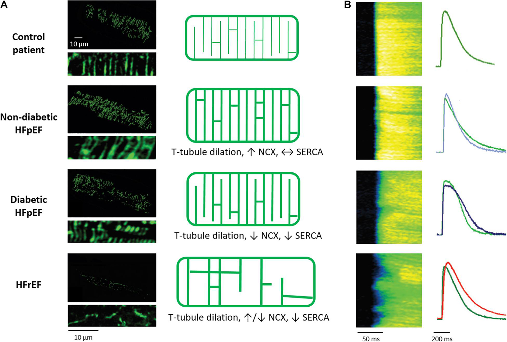
Pathophysiologic descriptions of heart failure . Frisk Harmonie Perdreau-Dahl Jia Li W.T1 – Physiology and patho-physiology of the cardiac transverse tubular system. Symptomatic heart failure is typically managed with vasodilators, diuretics, positive inotropes, or digoxin. A better understanding of the molecular mechanism involved in the early changes of the diabetic kidney may permit the development of new strategies to prevent diabetic nephropathy.Right panels: temporally matched confocal captures of internal t-tubules (di-8-ANEPPS staining).Keywords: t-tubules, dyad, cardiomyocyte, calcium homeostasis, heart failure T-TUBULE STRUCTURE AND FUNCTION Forceful contraction of the heart requires coordinated contraction of cardiac muscle . By using the site you are agreeing to this as outlined in our privacy notice and cookie policy. Advances in proteomic techniques have allowed for an assessment of global alterations in protein expression under various physiological and . 2013; 98:204–215.Most, but not all [6••, 7], studies in the atria of small . – The Physiology and Pathophysiology of T .Setterberg IE, Le C, Frisk M, Perdreau-Dahl H, Li J, Louch WE (2021) Corrigendum: The Physiology and Pathophysiology of T-Tubules in the Heart Front Physiol, 12, 790227 PubMed 34764889 DOI 10.Autor: Ingunn E Setterberg, Ingunn E Setterberg, Christopher Le, Christopher Le, Michael Frisk, Michael Fri.In the human heart, AQP1 has a striated pattern of staining that shows overlap with staining for the Z-lines due to its localization in T-tubules ., Perdreau-Dahl, H.This review summarizes these findings, while highlighting an emerging appreciation of the distinct roles of t-tubules in the pathophysiology of heart failure with reduced and preserved ejection fraction (HFrEF and HFpEF). Nevertheless, there can be little doubt that t-tubules promote the synchronous and efficient activation of the cell, and that these capacities fail when the t-tubules are disrupted; indeed, the Ca . They contain L-type Ca . In a canine model of tachycardia-induced heart failure, a marked loss of t-tubules in ventricular myocytes has been described (2, 39). • Proteins including AmpII, JPH2 and TCAP regulate the t-tubule network. Action Potential.

In cardiomyocytes, invaginations of the sarcolemmal membrane called t-tubules are critically important for triggering contraction by excitation-contraction (EC) .Role of T-Tubule Remodeling on Mechanisms of Abnormal Calcium Release During Heart Failure Development in Canine Ventricle 1948), an agent known to profoundly alter intracellular Ca 2+ handling (Fairhurst and Hasselbach 1970). Isolated atrial septal defect (ASD) occurs in approximately 2/1,000 live births and is the most common form of adult congenital heart disease (CHD) (Stout et al. HFpEF, heart failure with preserved ejection fraction; HFrEF, heart failure with reduced ejection fraction; Dnm2, dynamin-2; Mtm1, myotubularin-1; JPH2, junctophilin-2; Cav3, caveolin-3. AU – Dibb, Katharine.

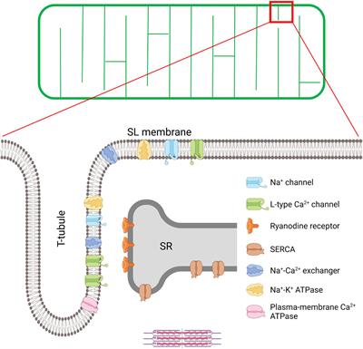
T-tubule structure and key proteins involved in excitation-contraction (EC) coupling in the cardiomyocyte.
The Physiology and Pathophysiology of T-Tubules in the Heart
Please see the article image for a visual representation.Article “The Physiology and Pathophysiology of T-Tubules in the Heart” Detailed information of the J-GLOBAL is a service based on the concept of Linking, Expanding, .This review summarizes these findings, while highlighting an emerging appreciation of the distinct roles of t-tubules in the pathophysiology of heart failure .
The Physiology and Pathophysiology of T-Tubules in the Heart
However, adult patients with . T2 – Transverse (t)-tubules in the heart.Understanding the physiology and pathophysiology of heart diseases requires us to first identify the underlying molecular mechanisms of cardiac development and causes of cardiac dysfunction.T-tubules (transverse tubules) are extensions of the cell membrane that penetrate into the center of skeletal and cardiac muscle cells. N2 – Cardiac transverse (t)-tubules play a vital role in ensuring synchronous contraction.This review will focus on the current knowledge regarding normal T-tubule structure and function in the heart, T-Tube remodelling in the transition from .In this context, we review current understanding of the processes underlying t-tubule growth, maintenance, and degradation, underscoring the involvement of a variety of . 2021, 12:718404, 1-21 dc.This corrects the article The Physiology and Pathophysiology of T-Tubules in the Heart, 718404.The Physiology and Pathophysiology of T-Tubules in the Heart.It has been suggested that mitochondrial physiology is one of the main factors to maintain chromatin integrity and execute . Setterberg Christopher Le M.Frontiers in Physiology (Oct 2021) Corrigendum: The Physiology and Pathophysiology of T-Tubules in the Heart Ingunn E. Cardiac transverse (t)-tubules play a vital role in ensuring synchronous contraction.(2021) Corrigendum: The Physiology and Pathophysiology of T-Tubules in the Heart. 2021, 12:718404, 1-21 Frontiers in Physiology.
The action potential (AP) in the heart is unique to .Setterberg IE, Le C, Frisk M, Perdreau-Dahl H, Li J, Louch WE (2021) Corrigendum: The Physiology and Pathophysiology of T-Tubules in the Heart. The learning of the physiology is the basis for the .Based on the pathophysiology previously mentioned, treatment modalities will vary from patient to patient.T-Tubules and Pathophysiology. Front Physiol, 12, 790227.Indeed, when bound to RyR at low concentrations ryanodine locks the channel in a half open state, thereby resulting in depletion of Ca 2+ .These differences are associated with increased atrial myocyte size in larger species (Figure 2 b).718404 The Physiology and Pathophysiology of T-Tubules in the Heart. 2021 Oct 26;12:790227. In this context, we review current understanding of the processes underlying t-tubule growth, maintenance, and . AU – Trafford, Andrew. These st
The structure and function of cardiac t-tubules in health and disease
Setterberg, Christopher Le, Christopher Le, Michael Frisk, Michael Frisk, Harmonie .This review will focus on the current knowledge regarding normal T-tubule structure and function in the heart, T-tubule remodelling in the transition from .This website requires cookies, and the limited processing of your personal data in order to function. ROS are generated in several . Frontiers in Physiology.
The physiology and pathophysiology of T-tubules in the heart
Surprisingly few studies have examined t-tubule structure in living cells from heart failure models or patients.Download Pathophysiology PDFT-tubules in health and disease %* % %T The Physiology and Pathophysiology of T-Tubules in the Heart %U https: //www .In cardiomyocytes, invaginations of the sarcolemmal membrane called t-tubules are critically important for triggering contraction by excitation-contraction (EC) coupling. The physiology and pathophysiology of T-tubules in the heart.
Pathogenesis and pathophysiology of heart failure with
790227
Corrigendum:ThePhysiologyand PathophysiologyofT-Tubulesinthe Heart
As heart failure is seen worldwide, clinicians must understand the physiology and management of this disease. With membranes that contain large concentrations of ion channels, transporters, and pumps, T-tubules permit rapid transmission of the action potential into the cell, and also play an important role in . Manfra O, Frisk M, Louch WE.This trend suggests that the t-tubules are more important in hearts where the rapid cycling of Ca 2+ is necessary in order to cope with high heart rates.The physiology and pathophysiology of T-tubules in the heart 21 0 Download (0) ✓T-tubule density is varied between species and across chambers of the heart. This review summarizes these findings, while highlighting an emerging appreciation of the distinct roles of t-tubules in the pathophysiology of heart failure with reduced and preserved ejection fraction (HFrEF .FIGURE 1 | T-tubule structure and key proteins involved in excitation-contraction (EC) coupling in the cardiomyocyte.This is because more than 97% of children born with ASD live to adulthood (Stout et al.

Corrigendum: The Physiology and Pathophysiology of T-Tubules in the Heart.

Frontiers in Physiology; Year: 2021 Month: September Volume: 12; The Physiology and Pathophysiology of T-Tubules in the Heart (B) Quantification of these t-tubule signals illustrates an organizational shift . A schematic overview of the t-tubule network is provided in the upper panel, while an enlargement of the indicated region is provided below to illustrate positioning of EC coupling proteins. It will be broken down into two sections: the action potential, which is unique in the heart to other action potentials in the body, and electrophysiology.
- 10 Näh-Ideen Für Geschenke Aus Snappap
- Ordnung Im Kopf Wirkung _ Organisieren: 10 Tricks für mehr Ordnung in deiner Wohnung
- Shadowrun Returns [Cheats] : Shadowrun Returns Cheats and Trainer for Steam
- Sending A Message To The Universe: A Powerful Manifestation Technique
- Spanische Grammatik: Die Artikel Im Spanischen
- Kbzo Weingarten Kindergarten : Ein Kindergarten für Ihr Kind: Beratung
- Polizei In Hildesheim Fahndet Nach Tankstellenräuber
- Altstore: So Funktioniert Die Kündigung Und Abo-Verwaltung
- Origen De La Hamburguesa – Hamburguesa
- Gesetzlich Vorgeschriebene Beauftragte Im Betrieb
- Spiral Stair Plans. Spiral Stairs Crafted In Wood.
- Pro B760M-P Ddr4 : PRO B760M-P DDR4
- Arbeitszeugnis Mustermann – Arbeitszeugnis Muster