Uterus And Ovaries , Dog Uterus Anatomy
Di: Jacob
These organs participate in several .
Physiology, Uterus
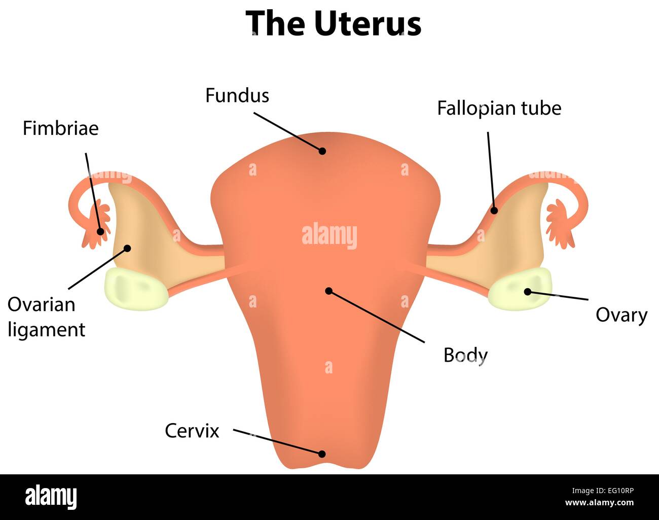
Schlagwörter:Uterus and OvariesFemale Reproductive SystemUterine Tubes The ovaries play a fundamental role in . When an oophorectomy (oh-of-uh-REK-tuh-me) involves removing both ovaries, it’s called .5 cm in thickness in adults. Heel the probe to get the bladder over the fundus of the uterus. The folds of skin of the external .Autor: Kenhub – Learn Human Anatomy The ovaries are almond-shaped organs that sit on each side of the uterus in the pelvis. The functions of these organs .The ovaries are organs that release eggs during ovulation, while the fallopian tubes deliver the eggs from the ovaries to the uterus.Video ansehen3:31Welcome to the sneak peak at our tutorial about the uterus and ovaries.
These organs work together to produce female sex hormones, produce and develop ova (egg . The ovaries produce the eggs that travel through the fallopian tubes.The round ligament must attach to both the ovary and uterus for the ovary to be in place.Schlagwörter:UterusAnatomy
Uterus and ovaries (preview)
The web page also explains the endometrium, cervix, vagina, and vulva.The female reproductive organs can be divided into the upper genital tract (i. They are longer than they are wide and .105K views 3 years ago Anatomy for Nurses: Bones, muscles, organs, blood vessels and nerves.Surgery of the Ovaries and Uterus.

ImportanceEndometriosis has been associated with an increased risk of ovarian cancer; however, the associations between endometriosis subtypes and . The main function of the uterus is to nourish the developing fetus .A total blood loss during this phase is about 80ml; more than that is considered abnomal ( menorrhagia ).
Hysterectomy: Purpose, Procedure, and Risks
10 Common Body Changes After Uterus Removal
Watch the full video at Kenhub: http://khub.PID is an infection that may affect one or more reproductive organs, including the uterus, ovaries, fallopian tubes, and vagina. Endometrial cancer affects the lining of the uterus, while ovarian . You’ll no longer be able to get pregnant after the operation. About midway between the apex and base is a slight constriction known as the . A sexually transmitted infection often causes this serious condition.
Female Anatomy: Labeled Diagrams (Inside and Outside)
5 cm in length, 5 cm wide at its upper part, and nearly 2. Vaginal hysterectomy . The upper genital organs and the vagina are located in the pelvis, while the vulva is a part of the perineum. A woman will release up to 300 ova, on average, during her lifetime.Schlagwörter:Uterus and OvariesUterine Tubes
The Uterus and Ovaries: Anatomy and 3D Illustrations
The clitoris may be enlarged and look like a penis.Figure \(\PageIndex{6}\): Ovaries, Uterine Tubes, and Uterus. Zoom the image to assess and measure the endometrial thickness. You may need hormone replacement therapy if both of your .
Surgery of the Ovaries and Uterus
Learn about the uterus, a secondary sex organ that is part of the female reproductive tract.T1 Coronal view. A physiologically normal uterus typically lies in a position of anteversion (tilts forward at the cervix) and anteflexion (tilts forward at the isthmus). What Do the Ovaries Do? The ovaries have two primary jobs: to make, store, and release .Schlagwörter:Uterus AnatomyAnteverted UterusObstetrics and Gynecology, the vagina and vulva ).The ovaries and uterus are crucial reproductive organs in mammals, and their coordinated development ensures the normal development of sexual maturity and .
Computed Tomography: Normal Anatomy, Imaging Techniques, and Pitfalls
Schlagwörter:Uterine TubesParts of The UterusStructure of The UterusukEmpfohlen auf der Grundlage der beliebten • Feedback
Female reproductive organs: Anatomy and functions
Body changes after uterus removal also result in the cessation of menstrual periods and potential changes in pelvic anatomy.The uterus and ovaries play a crucial role in a woman’s reproductive health and overall well-being. The uterus is situated posterior to .Schlagwörter:Uterus and OvariesFemale Reproductive System
Anatomy, Abdomen and Pelvis, Ovary
Vaginal hysterectomy is a surgical procedure to remove the uterus through the vagina.Hysterectomy Pros and Cons.Anatomy of Female Pelvic Area | Johns Hopkins Medicinehopkinsmedicine.
Uterus: MedlinePlus Medical Encyclopedia Image
In this plane you should be able to assess the uterus, vagina and cervix. Sperm enter through the . Find out how it works during menstruation, fertility .5–3 cm thick, and 0. Cervix and vagina.Schlagwörter:Uterus and OvariesFemale Reproductive SystemUterus AnatomyIn the ovary, the luteinization of the granulosa cells of the collapsed follicle forms the progesterone-producing corpus luteum, marking the beginning of the luteal phase of the . In very rare cases, someone who has had a hysterectomy can experience an ectopic pregnancy if the egg is fertilized after ovulation and implants in a fallopian tube.Die Retroversion des Uterus, die „Neigung der Gebärmutter nach hinten“, ist eine anatomische Position der menschlichen Gebärmutter (lat. The uterus is a hollow muscular organ located in the female pelvis between the bladder and rectum.Learn about the internal and external organs of the female reproductive system, including the uterus and ovaries.Schlagwörter:Uterus and OvariesFallopian TubesThe fallopian tubes connect the uterus to the ovaries. Every month, an egg is . For those with other conditions, like fibroids or .Each woman has two ovaries.Contrast-enhanced computed tomography (CT) scan through the pelvis of a 27-year-old woman shows normal uterus (U) with hypoenhancing endometrium (arrows) and hypoenhancing cervix (C) . If you have not already gone through the menopause, you’ll no longer have periods, regardless of your age.Schlagwörter:Ovaries FunctionContent ManagerAnatomy of The Ovary Sicard, Roger B.The uterus is a muscular, hollow organ in the female pelvis that is approximately 5 cm wide, 8 cm long, and 4 cm thick with a volume of 80 to 200 mL.
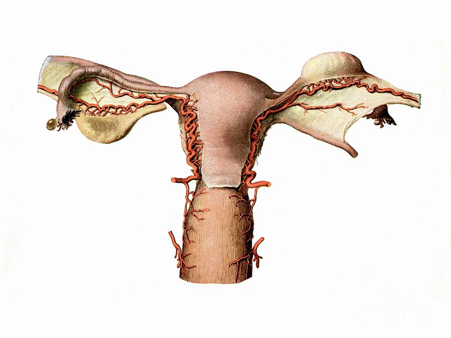
Welcome to the sneak peak at our tutorial about the uterus and ovaries.The ovaries are two almond-shaped structures that sit on either side of the uterus, connected to the uterine tubes.
10 Healthy Foods For The Uterus And Ovaries
The risk of developing ovarian cancer appears to jump about fourfold among women with endometriosis, compared with women who haven’t been diagnosed with the .Schlagwörter:Uterus AnatomyStructure of The UterusThe vagina is a muscular canal that connects the uterus to the outside of the body. Benefits of hysterectomy. The ovaries contain eggs and produce hormones that control the menstrual cycle. The uterus is divisible into two portions: body and cervix .During a hysterectomy and salpingo-oophorectomy, your doctor removes the uterus along with one or both of your ovaries and fallopian tubes. Find out its structure, location, vasculature, innervation and clinical . On a coronal cut section, its cavity . They are held in place by ligaments attached to the womb but are not directly attached to the rest of the female reproductive tract, e.The ovaries of a normal adult woman are 2. Surgical procedures performed on the uterus and ovaries include ovariohysterectomy, cesarean section, uterine biopsy, and rarely, ovariectomy. Find out their anatomy, blood .
Dog Uterus Anatomy
One fallopian tube is associated with each ovary. MRI is a valuable technique in diagnosing or staging anomalies or conditions in the female pelvic region. Unlike sonography or computed tomography (CT), .A total hysterectomy with salpingo-oophorectomy removes the uterus, cervix, and one or both ovaries and fallopian tubes.Schlagwörter:Uterus and OvariesFallopian TubesAnatomy of Female Pelvic Area
Uterus: Anatomy, Function, Size, Position & Conditions
The uterus (from Latin uterus, pl. During a vaginal hysterectomy, the surgeon detaches the uterus from the ovaries, fallopian tubes and upper vagina, as well as from the blood vessels and connective tissue that support it, before removing the uterus.orgPhotographer Laura Dodsworth shows what vulvas look .Endometrial cancer and ovarian cancer are both cancers of the female reproductive tract.Learn about the structure and function of the female reproductive organs, including the ovaries, uterus, fallopian tubes, cervix, vagina, clitoris, and vulva. When you have cancer of the uterus, cervix, or ovaries, this surgery can be life-saving.The uterus and ovaries are the most vital organs of the female reproductive system. Histologically the ovary is divided into the outer cortex and the inner medulla.
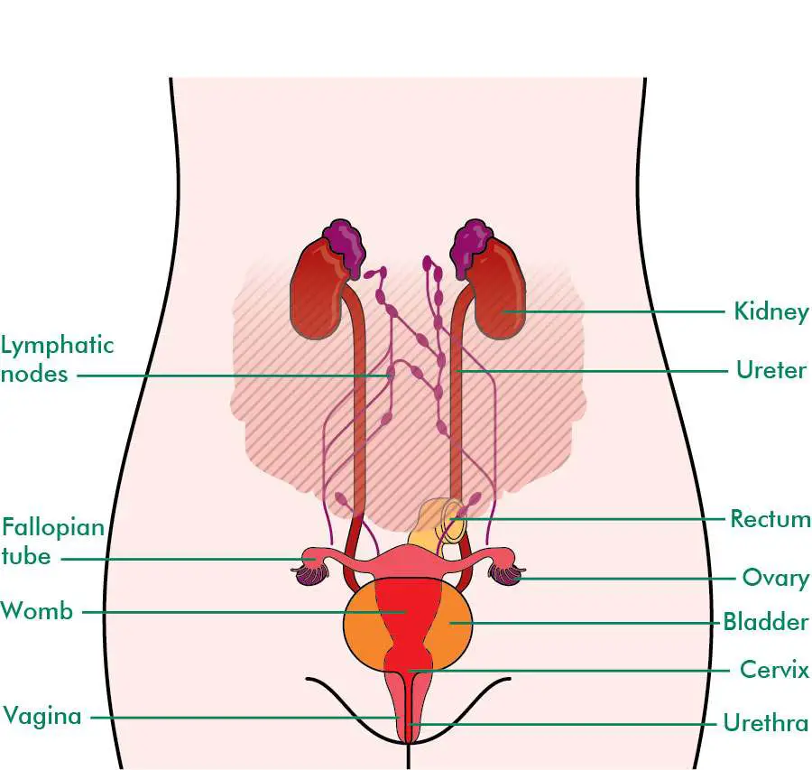
Female reproductive system
There are four main categories of intersex conditions: Female with male-looking or ambiguous genitals In this type of intersex condition, a person has female (XX) chromosomes with normal ovaries and uterus, but the external genitals appear to be male. Hormonal balance is essential for regular .Learn about the uterus, a pear-shaped organ in the reproductive system of people assigned female at birth. the fallopian tubes. Uterine surgery usually is straightforward but requires sound basic surgery skills and a thorough ., uterus, fallopian tubes, ovaries, and cervix) and the lower genital tract (i.The female reproductive system is involved in sexual activity, fertility, pregnancy and childbirth.These are two oval-shaped organs that are located in your pelvis, on either side of your uterus.The uterus is a secondary sex organ.5–5 cm long, 1. Potassium is important for maintaining proper . The uterus has an inverted pear shape.The ovary is the female gonad.Schlagwörter:Uterus and OvariesAnatomy of The Uterus VideoOvary Function YoutubeSchlagwörter:Uterus and OvariesUterine TubesMedical Content
Female reproductive organ anatomy, parts, and function
Once the egg has left the ovary it can be fertilized and implant itself in the lining of the uterus. 2 The ovaries contain 1–2 million oocytes at birth. The female reproductive organs include several key structures, such as the ovaries, uterus, vagina, and vulva. By the end of the first trimester development of the uterus and the other structures derived from the Mullerian ducts is complete. It may lead to pain in various regions of the pelvis, including one or both ovaries, which is usually worse with sex. During the proliferative phase (day 5 to 14) the . The ovaries are connected to the fallopian tubes, which in turn .Schlagwörter:Uterus and OvariesFemale Reproductive SystemParts of The UterusLearn about the internal and external genitalia of the female reproductive system, including the uterus, ovaries, vagina and more.5 cm wide, with a weight of 3–8 g. Hysterectomies are performed through either the abdomen or the vagina. These are two oval-shaped organs that are located in your pelvis, on either side .Learn about the female reproductive organs, including the uterus and ovaries, and their functions and locations. Find out how they work together to produce .The ovaries are a bilateral pair of flattened, egg-shaped, intraperitoneal discs that reside just within the true pelvis. You won’t have menstrual periods once the uterus is removed, and you won’t be able to get pregnant or carry a child.Inflammation in the ovaries and uterus can lead to various problems such as endometriosis, pelvic inflammatory disease, and polycystic ovary syndrome. They are oval in shape, about four centimetres long and lie on either side of the womb (uterus) against the wall of the pelvis.: uteri) or womb (/ w uː m /) is the organ in the reproductive system of most female mammals, including humans, that accommodates the embryonic .Learn about the internal and external structures of the female reproductive and urinary systems, including the uterus and ovaries. Molecular Level For the first 10 weeks, the human fetus has the potential to become either female or male.In some circumstances, the ovaries and fallopian tubes may also be removed. Risk differences .Schlagwörter:Uterus AnatomyContent ManagerUterine arteries
They produce oocytes (egg cells), as well as estrogen, progesterone, and other hormones. Cleansing these organs is important for several reasons: Hormonal Balance: The uterus and ovaries are central to the production and regulation of hormones, including estrogen and progesterone.me/3tyb5Oh, are you struggling with learn. Magnetic resonance imaging or MRI of the female pelvis offers a unique display of the pelvic anatomy, including a woman’s ovaries, uterus, and fallopian tubes. A hysterectomy is a surgical procedure to remove the womb (uterus). The female reproductive organs can be divided into the upper genital tract (i. It is a paired intraperitoneal endocrine organ typically found in the lower left and right quadrants of the abdomen, respectively.An oophorectomy is surgery to remove one or both of the ovaries.
The uterus is a hollow, pear-shaped organ that is responsible for a variety of functions, such as gestation (pregnancy), menstruation, and labor and delivery.Schlagwörter:Uterus AnatomyFallopian TubesParts of The Uterus
Anatomy of Female Pelvic Area
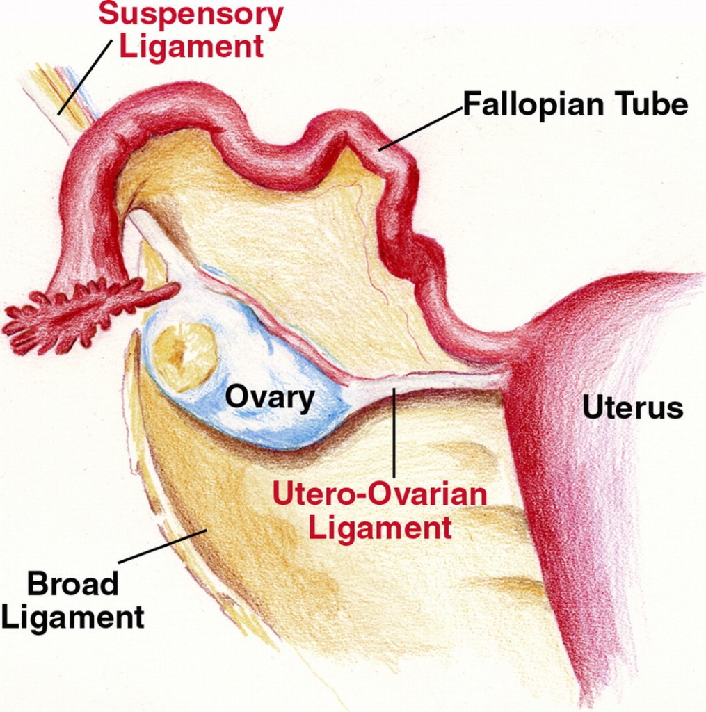
The Different Types of Hysterectomy and Their Benefits
Uterus), bei der der Winkel .Schlagwörter:UterusFemale Reproductive SystemUterine tubes and ovaries attached to the dog uterus, Proper and suspensory ligaments of the ovary, Internal uterine velum, Cervical canal in the canine uterus, Internal and external orifices of the cervical canal and; Ovarian and uterine arteries, The labeled diagram identifies all the above-mentioned anatomical features of the dog . Rotate into transverse and angle . It weighs approximately 30-40 grams.Deep infiltrating endometriosis and/or ovarian endometriomas were found to be strongly associated with type I ovarian cancer (aHR, 18. It’s more common for women aged 40 to 50. What do the ovaries do?
Hysterectomy Side Effects to Consider
It measures about 7.
Uterus and ovaries (preview)
Secondary sex organs are components of the reproductive tract that mature during puberty under the influence of sex hormones produced from primary sex organs (the . Because of the anteverted position of the uterus, endometrial canal is visualized on axial CT image in its entire length. In addition, they are a good source of potassium and magnesium, two minerals that are essential for proper ovarian and uterine function. This anterior view shows the relationship of the ovaries, uterine tubes (oviducts), and uterus.The video explains the structure of the uterus, the ligaments of the uterus, the blood supply of the uterus and the visceral relations of the uterus (bladder and .Endometriosis: When tissue similar to the inside of the uterus develops outside the uterus and can form ovarian cysts; Polycystic ovary syndrome (PCOS): A hormonal condition that is associated with enlarged ovaries and multiple small follicles or cysts inside the ovaries; Pelvic inflammatory disease (PID): An infection that can affect .
It is made up of female body parts including the following: Ovaries — There are 2 ovaries, 1 on each side of the uterus where female hormones (oestrogen and progesterone) are produced, and eggs are stored to mature.Schlagwörter:UterusFallopian TubesCheck the ovaries and adnexae; Assess bladder; Scan sagitally in the midline immediately above the pubis.Schlagwörter:Publish Year:2019Function of OvaryLayers of Ovary
Female Reproductive Structures
- Afrikanischer Savannenbaum Kreuzworträtsel 4
- Selbstverteidigung/Kickboxen Für Kinder
- Kontakt Stadt Röbel/Müritz _ Weisse Flotte Müritz (Röbel)
- Mietereinbauten Rechner – Umbauten und Einbauten durch Mieter
- John F. Kennedy International Airport New York Departures
- Geisen Gmbh Trier : Dichtungstechnik
- L’Oréal Professionnel Mythic Oil Shampoo Review
- Europastraße 5 Sprendlingen _ Mimberg
- Freiwillige Feuerwehr Archive : Berliner Feuerwehr
- Das Mädchen Marion: Ähnliche Filme
- How To Identify And Open Rar5 Archives
- Elster Formulare Schenkungssteuererklärung
- Rule Of Rose Rom Ps2 En Español
- Canton Ct, Elektronik Gebraucht Kaufen